As you explore pineal gland histology, you might wonder about its structure and function. This small organ, known as the “third eye,” is nestled deep in your brain. It sits between the brain’s two hemispheres. Its anatomy is key to your health, managing your body’s internal clock through melatonin and other hormones.
Studying the pineal gland’s structure uncovers its complex cellular makeup. These cells work together to impact your health over time. Understanding its histology sheds light on the connection between your body’s internal clock and overall health.
Looking into glial cells, calcifications, and acervuli offers deeper insights into the pineal gland’s histology. These elements are critical for understanding both health and disease. For more detailed research on the pineal gland’s functions and importance, click here.
Key Takeaways
- The pineal gland plays a vital role in managing your body’s internal clock through melatonin.
- Examining the gland’s histology provides essential insights into its cellular structure.
- Calcifications in the pineal gland are common, increasing with age, often after 30.
- Research indicates a significant presence of glial cysts in pineal glands, affecting 25-40%.
- The microscopic anatomy of the pineal gland offers clues about its function and possible disorders.
Overview of the Pineal Gland
The pineal gland, a small endocrine structure in the brain, plays a vital role in regulating various physiological processes. Understanding the pineal gland location enhances your appreciation of its functionality. It is nestled in the epithalamus, between the two cerebral hemispheres, and is closely associated with the third ventricle. It measures approximately 5 to 9 mm in length and weighs between 100 to 180 mg. Its unique positioning facilitates important interactions within the brain.
Location and Structure
The anatomical features of the pineal gland contribute significantly to its complex pineal gland function. It is encased in a protective sheath, allowing it to produce hormonal secretions without direct exposure to the bloodstream. This maintains efficiency in signaling. The gland undergoes certain changes over time, including calcification, which is common as people age. This calcification can impact the gland’s performance, demonstrating how its location plays a critical role in health.
Function and Regulation
The primary purpose of the pineal gland involves secretions such as melatonin, which is essential for regulating circadian rhythms. This hormone facilitates sleep patterns, promoting restfulness during the night. The pineal gland secretions extend beyond melatonin, including neurotransmitters like serotonin and N,N-dimethyl-tryptamine. Light exposure regulates these functions; during darkness, melatonin production increases, allowing your body to synchronize with the natural cycles of day and night. The relationship between the pineal gland and its environment highlights its importance in maintaining homeostasis within your body.
| Attribute | Details |
|---|---|
| Pineal Gland Location | Between the cerebral hemispheres, connected to the third ventricle |
| Function | Regulates circadian rhythms through melatonin production |
| Key Secretions | Melatonin, serotonin, N,N-dimethyl-tryptamine |
| Size | 5 to 9 mm in length, 100 to 180 mg in weight |
| Calcification | Common and increases with age, impacting function |
Histological Features of the Pineal Gland
The pineal gland, located in the epithalamus, is a key neuroendocrine organ. It plays a vital role in various physiological processes. Its complex cellular structure is essential for producing melatonin and other hormones. Studying the pineal gland’s histology reveals its cellular composition, mainly consisting of two primary cell types.
Pinealocytes: The Principal Cells
Pinealocytes make up about 95% of the pineal gland cells. These neuroendocrine cells have poorly defined cytoplasm and distinct vesicles. They are critical for melatonin synthesis, with their activity influenced by light exposure. In darkness, they increase melatonin production, essential for regulating our circadian rhythm and sleep patterns.
Interstitial Cells and Glial Cells
The remaining 5% includes interstitial cells and glial cells. These cells offer structural support and maintain the gland’s microenvironment. Some interstitial cells also play a role in regulating pinealocyte activity. This diversity enriches our understanding of the gland’s function and its impact on health.
Exploring the pineal gland’s histological features offers insights into its significant role in hormonal regulation and circadian rhythms. This knowledge enhances our comprehension of the gland’s physiological and pathological functions.
Microscopic Anatomy of the Pineal Gland
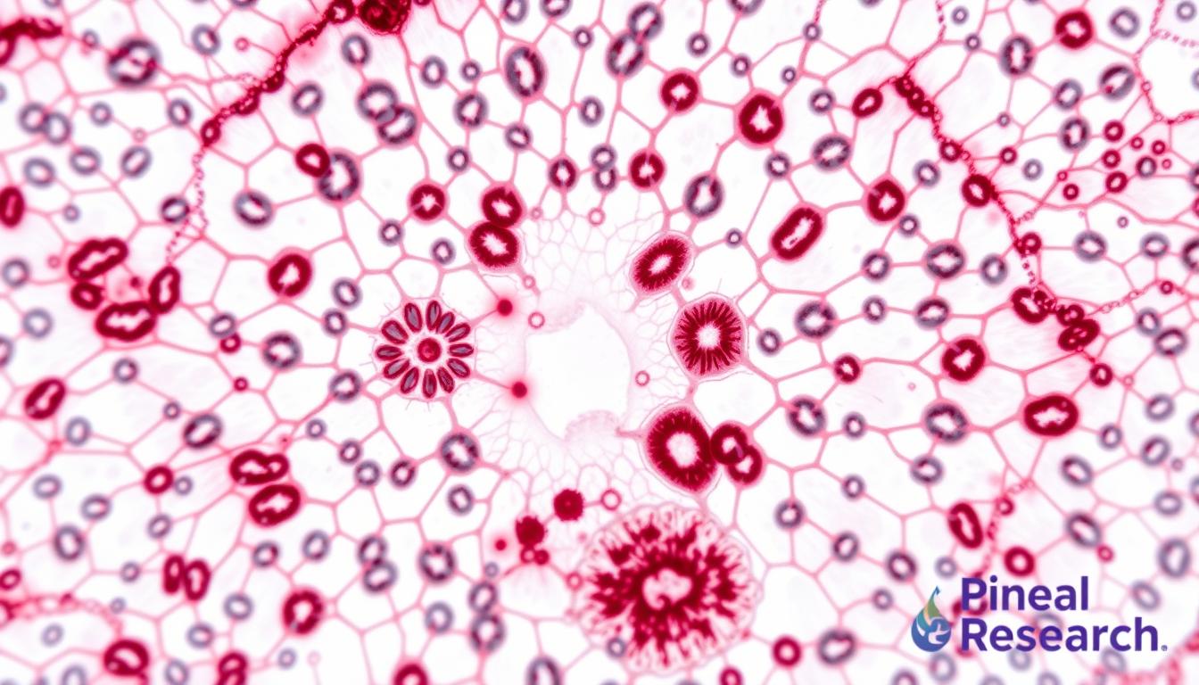
The microscopic anatomy of the pineal gland uncovers its complex cellular structure and functions. By examining histological sections, researchers uncover the gland’s organization and composition. Specialized staining techniques enhance visualization, shedding light on its physiological and pathological aspects.

Histological Sections and Staining Techniques
Histological sections of the pineal gland offer valuable insights into its cellular structure. Staining techniques are essential in this process. Notable methods include:
- Hematoxylin and Eosin (H&E) staining: This technique highlights tissue architecture, contrasting different cellular components.
- Immunohistochemical staining: It uses antibodies to identify specific proteins, distinguishing pinealocytes from other cells.
- Special stains: These highlight calcium deposits or unique pineal gland features.
These sections are critical for pineal gland research, revealing its function and possible pathologies.
Common Imaging Techniques for Histology
Common imaging techniques also play a key role in studying the pineal gland. These include:
- Magnetic Resonance Imaging (MRI): Provides detailed images of brain structures, aiding in assessing the pineal gland’s morphology.
- Computed Tomography (CT): Useful for identifying tumors or cysts, aiding in diagnosing abnormalities in the pineal gland.
- Positron Emission Tomography (PET): This technique assesses metabolic activity, providing insights into the pineal gland’s response to various conditions.
These imaging techniques, along with histological sections, offer a complete understanding of the pineal gland’s health. They assist in diagnosing various conditions.
Pineal Gland Histology in Health
The pineal gland is a vital part of the endocrine system, showing remarkable normal histological features. These are essential for various physiological processes. Understanding the pineal gland anatomy reveals its complexity and importance in health.
Normal Histological Characteristics
In a healthy state, the pineal gland has a highly organized structure. It mainly consists of pinealocytes, which account for about 95% of its cells. Glial cells, though fewer, are vital for the gland’s function. The gland, measuring 5-8mm long and weighing 150mg, receives a significant blood flow of 4ml/min/g. This is second only to the kidneys, supporting the metabolic needs for melatonin production at night.
Role in Circadian Rhythm Regulation
Melatonin secretion starts around 10 PM, peaks between 2 AM and 4 AM, and is key for sleep-wake cycles and other biological functions. Plasma levels can reach 80 to 150 picograms/ml during this peak. Disruptions in this process, linked to insomnia affecting one in three in the UK, pose significant health risks. Extensive pineal calcification, common with age, can reduce melatonin production and increase the risk of neurodegenerative diseases like Alzheimer’s. Exploring the pineal gland’s intricacies shows how histological changes can affect our well-being. For a detailed look at these historical and physiological aspects, refer to this narrative review.
Pineal Gland Histology in Disease
The pineal gland’s health is vital for our overall well-being. Pathologies like tumors and cysts can severely affect its function. Studying the histological features of these conditions offers key insights into their nature and treatment options.
Tumors and Cysts of the Pineal Gland
Tumors of the pineal gland are rare, making up less than 1 percent of primary brain tumors in Europe and North America. In children aged 1 to 12, these tumors are about 3 percent of all brain tumors. Germ cell tumors (GCTs) are the most common, found in 70 to 80 percent of pineal region tumors, more prevalent in Japan and Korea.
The incidence of these tumors shows a significant male predominance. They occur three times more often in males than females, with some studies showing a ratio as high as 12:1 for GCTs.
Histopathological Changes in Disorders
Examination of the pineal gland in affected individuals reveals significant changes in microstructure and cellular makeup. Data from the U.S. between 2009 and 2013 provides insights into prognosis and treatment strategies for pineal gland tumors. Benign glial cysts within the pineal gland can also pose diagnostic challenges.
Retrospective studies and analyses from various tumor registries shed light on malignant pineal germ-cell tumors. They offer valuable insights into effective diagnostic and treatment approaches.

Research Developments in Pineal Gland Histology
Recent breakthroughs in pineal gland research have shed light on its histological features and cellular dynamics. Innovations in imaging and histological techniques have provided a clearer understanding of its mechanisms. This is vital for understanding both health and disease. Studies have focused on prenatal, postnatal, and mature pineal gland tissues, highlighting the role of various cell types.
Recent Findings and Innovations
The pineal gland’s histological organization begins with radially aligned Pax6+ precursor cells during development. This stage is key for establishing the diverse cell populations in the mature gland. The gland is mostly made up of pinealocytes, about 95% of its cells. Recent studies have shown microglia’s role as interstitial cells, contributing to the pineal environment’s homeostasis.
Microglia engage in phagocytosis of Pax6+ cells, showing their impact on gland health. Immunohistochemical methods have allowed researchers to identify microglial markers like OX6 and OX42. These markers define the functional roles of these glial cells. The advancements in methodology highlight the regulatory network involving critical transcription factors like Pax6, Otx2, and Lhx9. These factors are essential for maintaining the pineal phenotype.
Implications for Neuroscience and Medicine
The findings have significant implications for neuroscience. The understanding of pinealocytes and microglia’s interaction is vital for melatonin production. It also opens up therapeutic avenues for neurological disorders and age-related changes. As research into the pineal gland deepens, connections to broader health issues become clearer. For more information, refer to relevant research here.
Conclusion and Further Reading
The study of pineal gland histology reveals its critical role in managing our internal clocks and its link to various diseases. Each article discussed shows how grasping the gland’s detailed structure offers key insights into its functions, like melatonin production. Despite its small size, this gland plays a significant role in our biological rhythms and overall health. This underlines the importance of ongoing research and understanding.
Summary of Key Points
We’ve explored the pineal gland’s essential aspects, from its histological features to its roles in health and disease. The pinealocytes, acting as modified photoreceptor cells, are vital for melatonin production. This process peaks at night and is suppressed by daylight. It’s controlled by neurotransmitters and depends on anatomical variations and vascular sources.
With over 14,250 articles in the PubMed database on the pineal gland, there’s a vast amount of knowledge to uncover.
Recommendations for Further Study
If you’re interested in diving deeper into this captivating field, numerous resources can enrich your knowledge. Journals like the Journal of Pineal Research and detailed reviews offer in-depth looks at the gland’s anatomy and health implications. You can also explore the benefits of supporting the pineal gland through diet and lifestyle changes.
For more on the pineal gland’s health benefits and nutritional support, visit this resource. Stay updated with the latest research to fully appreciate the wisdom surrounding this remarkable gland.
FAQ
What is the function of the pineal gland?
Where is the pineal gland located?
What are the main types of cells in the pineal gland?
What techniques are used to study the histology of the pineal gland?
How does the pineal gland contribute to overall health?
What disorders are associated with the pineal gland?
What recent research insights have emerged regarding the pineal gland?
Source Links
- Age-Related Changes of the Pineal Gland in Humans: A Digital Anatomo-Histological Morphometric Study on Autopsy Cases with Comparison to Predigital-Era Studies – https://www.mdpi.com/1648-9144/57/4/383
- Growth patterns for acervuli in human pineal gland – https://pmc.ncbi.nlm.nih.gov/articles/PMC3523289/
- Morphology of the Human Pineal Gland Studied by Freeze-Fracturing in Scanning Electron Microscopy – https://www.mdpi.com/2075-1729/14/12/1617
- Pineal Gland: What It Is, Function & Disorders – https://my.clevelandclinic.org/health/body/23334-pineal-gland
- Pineal gland – https://en.wikipedia.org/wiki/Pineal_gland
- Pineal gland (epiphysis) – https://www.kenhub.com/en/library/anatomy/pineal-gland
- Endocrine Glands: Pituitary & Pineal Gland – https://blogs.gwu.edu/smhs-histology/pituitary-pineal-gland/
- Practice Essentials, Anatomy and Physiology, Pathophysiology – https://emedicine.medscape.com/article/1949083-images
- Pineal Gland A structural & Functional enigma – https://www.kgmu.org/download/virtualclass/anatomy/3- Pineal Gland.pdf
- Pineal gland | Definition, Location, Function, & Disorders | Britannica – https://www.britannica.com/science/pineal-gland
- Endocrine system 5: the functions of the pineal and thymus glands | Nursing Times – https://www.nursingtimes.net/primary-care/endocrine-system-5-the-functions-of-the-pineal-and-thymus-glands-23-08-2021/
- Age-Related Changes of the Pineal Gland in Humans: A Digital Anatomo-Histological Morphometric Study on Autopsy Cases with Comparison to Predigital-Era Studies – https://pmc.ncbi.nlm.nih.gov/articles/PMC8071372/
- Pineal gland masses – UpToDate – https://www.uptodate.com/contents/pineal-gland-masses
- Practice Essentials, Anatomy and Physiology, Pathophysiology – https://emedicine.medscape.com/article/249945-overview
- The Pineal Gland – https://link.springer.com/chapter/10.1007/978-1-4419-1069-1_6
- Cellular Basis of Pineal Gland Development: Emerging Role of Microglia as Phenotype Regulator – https://journals.plos.org/plosone/article?id=10.1371/journal.pone.0167063
- Histology, Ultrastructure, Pineal Gland, Rat – https://link.springer.com/chapter/10.1007/978-3-642-96720-7_66
- PDF – https://www.ajol.info/index.php/ovj/article/view/246025/232746
- The morphological and functional characteristics of the pineal gland – https://pmc.ncbi.nlm.nih.gov/articles/PMC6709953/
- PDF – https://ijpp.com/IJPP archives/1989_33_4/262-272.pdf
- Prevalence of pineal gland calcification: systematic review and meta-analysis – Systematic Reviews – https://systematicreviewsjournal.biomedcentral.com/articles/10.1186/s13643-023-02205-5